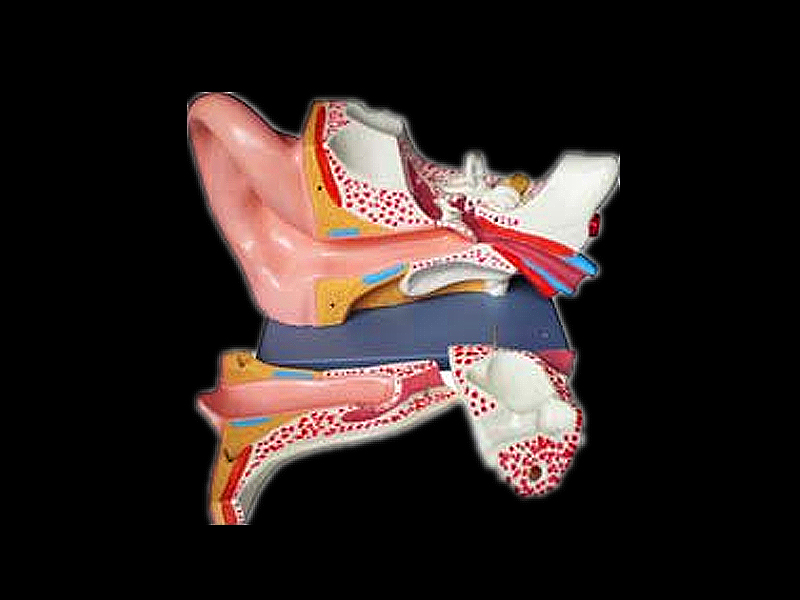

The lateral surface is covered by a thin layer of skin, which is formed mainly by the perichondrium and an extremely fine subcutaneous layer. It is divided into two surfaces: lateral and medial. The auricle is the lateral-most part of the external ear and is composed mainly of cartilage and skin.

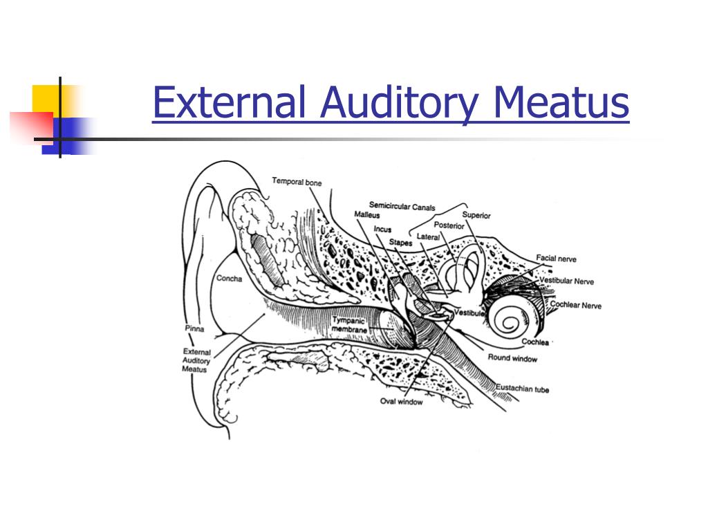
This part is closely related to the anterior and posterior canal for the chorda tympani and the petrotympanic suture. The tympanic membrane within the notch of Rivinus, above the anterior and posterior malleolar folds extending to the lateral process of the malleus, is called the pars flaccida. Above the superior end of the tympanic sulcus, the tympanic annulus becomes a fibrous band. The edge of the tympanic membrane is thickened to form a fibrocartilaginous ring (tympanic annulus) attached to the tympanic sulcus, an incomplete ring of the tympanic bone that is interrupted by the notch of Rivinus. The tympanosquamous suture runs between the postglenoid process and the tympanic part of the temporal bone. At the anterior edge of this roof, the postglenoid process is positioned between the mandibular fossa and anterior wall of the external acoustic meatus. The posterior root of the zygomatic arch of the squamous part forms the roof of the external acoustic meatus. The endomeatal spine is formed by a projection of the tympanosquamous suture into the canal. The suprameatal spine (Henle’s spine) is situated at the upper and posterior part of the orifice of the external acoustic meatus. Additionally, two spines are identified in the bony portion of the meatus: endomeatal and suprameatal spines. The tympanosquamous suture, between the squamous and tympanic parts, is continuous medially with the petrotympanic and petrosquamous fissures ( Fig. The tympanic part produces three sutures: tympanosquamous, tympanomastoid, and petrotympanic. The superior and posterior bony walls are formed by the squamous and mastoid parts, and the squamomastoid suture can be identified ( Fig. The anterior and inferior bony walls are formed by the tympanic part. The bony canal of the external acoustic meatus is composed of three parts of the temporal bone: squamous, tympanic, and mastoid.

The temporal bone consists of five components: squamous, tympanic, petrous, mastoid, and styloid parts ( Fig. Finally, there is a short description of clinical considerations. This chapter examines the microsurgical anatomy of the auricle and external acoustic meatus in cadaveric dissections and organizes the results in the following sections: (1) bone structure, (2) auricle, (3) cartilaginous skeleton of the auricle, (4) external acoustic meatus, (5) muscles, (6) neural innervation, (7) vascular supply, and (8) fascial structure. Surgery resulting in cosmetic and functional satisfaction relies not only on the surgeon’s skill and use of advanced technologies but also on their complete understanding of the area’s microsurgical anatomy, which makes surgery gentler and safer. The external ear is a focus of otologic and plastic surgery, but it is also important in neurologic and lateral skull-base surgery. It is also cosmetically important and its anatomical structures are extremely complicated and delicate. The auricle is a concave structure that directs sound waves into the external acoustic meatus. The external ear is formed by the auricle, external acoustic meatus, and tympanic membrane. Noritaka Komune, Junichi Fukushima, and Albert L.


 0 kommentar(er)
